medical innovation
How Cryo-Electron Microscopy (Cryo-EM) is revealing the invisible?
Cryo-EM is an innovation heralding a transformational shift in how researchers perceive and understand microscopic phenomena.
Innovation and automation has always been the critical components driving scientific progress forward. Nonetheless, some particularly significant advances have arisen due to rapid technological advancements in recent years. One example lies with the rise of cryo-electron microscopy (cryo-EM) - an innovation heralding a transformational shift in how researchers perceive and understand microscopic phenomena.
Cryo-EM captures images showing biological molecules existing within their native state. These unique images offer unrivalled insights into molecular structure and function, never before achievable by other means. This article investigates how this cutting-edge technology reveals elements once believed invisible - while also exploring its potential for inspiring further innovations within the field.
What is Cryo-Electron Microscopy?
The incredible technique known ascryo-electron microscopy (cryo-EM) has revolutionized our ability to observe biological molecules at high resolutions. Essentially, this method involves flash-freezing specimens in an ultra-thin layer of ice, which hardens around them. Electrons are then beamed onto these samples; they interact with various parts of the molecules under study before being captured by detectors, which produce two-dimensional images. By taking multiple snapshots from different angles and using advanced mathematical algorithms to stitch them together into three-dimensional models, scientists can recreate incredibly detailed structures with amazing accuracy.
Cryo-EM was initially limited by low-resolution levels and demanding requirements such as highly purified samples when it was first pioneered in the 1970s. In recent years, significant technological improvements have made it one of the most powerful tools available for structural biologists.
Cryo-electron microscopy (cryo-EM) has been pivotal in producing exceptionally sharp images of diverse biological molecules such as viruses, membrane proteins, or even whole cells at unprecedented resolutions.
How Cryo-EM is revolutionizing structural biology
The progress made in recent years in the field of structural biology owes much to advanced technologies such as cryogenic electron microscopy (cryo-EM). By providing images of biological molecules in their native state, cryo-EM has revolutionized how researchers view and understand the microscopic world. Such insights into molecular structure and behaviour have led to significant discoveries with implications ranging from new treatments for illnesses to fundamental new paradigms for scientists working with these materials.
One major advantage offered by this technique is its ability to examine biomolecules while they are situated naturally within an organism rather than in isolated samples. Among other reasons, this quality makes cryo-EM a preferred method when studying large, complex molecules like proteins or enzymes that are extremely difficult to analyse otherwise. Unique information about protein conformational flexibility, as it moves within its environment, can be obtained through the use of cryo-EM methods effectively employed during interaction analysis with various components.
Cryo-electron microscopy (cryo-EM) has been pivotal in producing exceptionallysharp images of diverse biological molecules such as viruses, membrane proteins, or even whole cells at unprecedented resolutions. As a result, insights into their function and structure are being discovered at an accelerating pace never seen before.
Insights into their function and structure are being discovered at an accelerating pace never seen before.
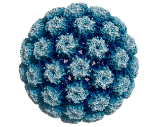
Color-enhanced scanning electron micrograph, See page for author, Public domain, via Wikimedia Commons
For instance, cryo-EM has been employed in investigating the HIV structure, leading to innovative drug discovery aimed at eradicating it from patients' bodies. Alzheimer's-associated protein structure information obtained from similar studies aims to identify newer and better treatment methodologies for this disease, making it an exciting period for research altogether!
Another marquee feature is cryo-EM's capability of snapping a picture of a molecule present in various conformations since many biomolecules exhibit dynamic behaviour, with each conformation having different functional significance, providing immense potential for scientists worldwide to further our understanding of molecular-level structures and functions. These are critical markers for advancing the biological sciences.
By providing images of molecules in their natural state, cryo-EM is enabling researchers to gain greater insight into how these entities operate within the complex workings of the human body.
The potential for further innovation
Despite impressive success thus far in cryo-EM research, opportunities for continued growth abound. Special attention is presently devoted toautomating aspects of the cryo-EM process to improve sample throughput for efficient analysis en masse. Robotics stands as one particular avenue being developed toward this end-point. By handling samples more accurately than humans can, robots limit contamination risks while simultaneously minimizing human-generated errors - advancing bothreproducibility and efficiency alike.
Beyond this promising direction in automation lies research into innovative imaging techniques specifically designed to enhance cryo-EM capabilities. Work on new detectors continues apace in hopes that these technological improvements may lead to capturing high-resolution images with greater speed. Innovative advancements have recently been made within the arena of cryo-electron microscopy through the introduction ofdirect electron detectors.
These new devices boast a quicker image-capturing ability than previous film-based methods leading to maximized efficiency during research and data collection processes. This improvement bodes well for scientists seeking valuable insights through cryo-EM analysis.
Scientists are actively investigating how best to combine different approaches including X-ray crystallography and nuclear magnetic resonance (NMR) spectroscopy, as complementary methods for forming a holistic perspective concerning biological molecules.
The study of novel approaches aimed at visualizing large molecular complexes presents another active area of research interest in contemporary times, with techniques such aselectron tomography at the forefront. This methodology enables researchers to obtain a three-dimensional account of complex molecular structures, providing critical insights into their behaviour and functionality. Notwithstanding its promise, it remains relatively nascent in terms of adoption by researchers across disciplines necessitating ongoing refinement efforts for enhanced uptake among scientific communities worldwide.
Furthermore, cryo-EM employed alone may have limitations; hence scientists are actively investigating how best tocombine different approaches including X-ray crystallography and nuclear magnetic resonance (NMR) spectroscopy, ascomplementary methods for forming a holistic perspective concerning biological molecules. Each method offers unique benefits. Cryo-EM, for instance, provides high-resolution information about molecular shape, while X-ray crystallography pinpoints atomic positions in certain molecules. There have been exceptional strides made through cryo-EM in structural biology, leading to previously unearthed knowledge on biomolecules.
In summary, as technology continues to improve, the limits of research possibilities concerning biological entities will continue to expand. Scholars are currently investigating novel approaches to automating the cryo-EM procedure. They aspire to develop advanced detectors that can capture higher-quality images at faster rates. Furthermore, they plan to integrate cryo-EM with other techniques to obtain a fuller depiction of biological molecules. These advances will continue to drive scientific progress and innovation in the years to come.
Conclusion: Cryo-EM is unlocking the secrets of life and fuelling scientific progress
In conclusion, structural biologists hold cryo-electron microscopy (cryo-EM) in high regard as an influential instrument at their disposal. It empowers them with a remarkable understanding of how biological molecules are structured and operate within living organisms.
Utilizing this technique has already yielded valuable findings that have improved treatment options for numerous diseases globally, establishing potential growth in research endeavours with every advancement made possible by its evolution over time. As such, cryo-EM's role becomes ever more prominent in driving forward scientific progress toward discoveries that could change our world profoundly.
Color-enhanced scanning electron micrograph, See page for author, Public domain, via Wikimedia Commons


Share this article


